Mesothelioma Chest Pain: How It Differs from Cardiac, Pleural, and Muscular Causes
Mesothelioma chest pain is usually dull, lateralized, and persistent. How it differs from cardiac, pleural, and chest wall causes.
Chest pain is one of the two symptoms that most often brings someone with mesothelioma to a doctor for the first time. The other is shortness of breath. The problem is that chest pain in a 65-year-old former insulator looks, on the surface, like a dozen other things: a heart problem, a stubborn pneumonia, a rib strain from yardwork. The clinical work of separating them is what determines how quickly the diagnosis gets made.
This guide walks through what the primary clinical literature says about the character of mesothelioma chest pain, and how it differs from the alternatives a clinician will weigh on the same visit. It draws on the open-access European Respiratory Review update by Bibby and colleagues, the National Cancer Institute Mesothelioma Treatment (PDQ), the 2021 AHA/ACC chest pain guideline, and the American Family Physician review of costochondritis.
What Mesothelioma Chest Pain Feels Like
The 2016 European Respiratory Review update on malignant pleural mesothelioma, by Bibby and colleagues, describes the typical character in plain language: chest pain in mesothelioma “is usually dull and heavy and sometimes described as a ‘dragging’ sensation.” The pain can come from the pleural effusion, from the tumor itself, or from both. Pleuritic pain, meaning sharp pain that worsens with each breath, is less common, although it does occur when there is parietal pleural irritation.
That description sets mesothelioma chest pain apart from the textbook patterns of other things on the differential. It is not the central crushing pressure of cardiac angina. It is not the sharp, breath-locked stab of acute pleurisy. It is not the focal, point-tender ache of costochondritis. It is duller, more constant, and tied to one side of the chest.
A few features recur in the case literature:
- The pain is usually unilateral, on the side of the tumor and effusion.
- It is persistent rather than intermittent, and tends not to be relieved by rest, position changes, or short courses of NSAIDs.
- It can refer to the shoulder on the same side when the diaphragmatic pleura is involved, because the diaphragm shares innervation (the phrenic nerve) with the shoulder.
- It worsens with disease progression, especially when the tumor begins to invade the chest wall.
Bibby and colleagues note that chest pain “tends to worsen as the disease progresses, particularly if invasion of the chest wall occurs,” and that “bone pain secondary to rib invasion or neuropathic pain from intercostal nerve involvement may also feature.” That trajectory, from a vague unilateral ache to a fixed, severe pain with neuropathic features, is one of the more useful pattern recognitions for clinicians and families.
Why a Pleural Effusion Drives So Many of the Symptoms
In early-stage disease, the most common reason a person with mesothelioma feels worse is fluid: a pleural effusion that compresses the lung on the affected side. Bibby et al report a pleural effusion at presentation in roughly 70% of patients, and the NCI’s Mesothelioma Treatment (PDQ) Health Professional Version states that pleural and peritoneal effusions are major symptomatic problems for at least 66% of patients overall.
A unilateral effusion does two things at once. It compresses underlying lung tissue, producing the shortness of breath that often dominates the early presentation. And it stretches the parietal pleura, which is densely innervated and is the source of most of the chest discomfort at this stage. Drainage (thoracentesis) produces brief relief, the fluid recurs, and the cycle prompts the workup that leads to a diagnosis.
A recurrent unilateral pleural effusion in someone with documented asbestos exposure is one of the strongest signals on the differential. It is not, by itself, mesothelioma. But it is the finding that should put mesothelioma firmly on the list alongside the other causes of exudative effusion any thoracic team will work through.
How Cardiac Chest Pain Differs
The 2021 American Heart Association/American College of Cardiology Guideline for the Evaluation and Diagnosis of Chest Pain (Gulati et al, Circulation) describes ischemic cardiac pain as characteristically “deep, difficult to localize, and usually diffuse.” The guideline lists pain, pressure, tightness, or discomfort in the chest, shoulders, arms, neck, back, upper abdomen, or jaw, alongside shortness of breath and fatigue, as anginal equivalents.
Several features push a clinician away from cardiac ischemia and toward something else. The guideline puts it directly:
“Pain described as sharp, fleeting, related to inspiration (pleuritic) or position, or shifting locations suggests a lower likelihood of ischemia. … Chest tenderness on palpation or pain with inspiration markedly reduce the likelihood of ACS.”
The contrast is fairly clean. Mesothelioma pain is unilateral and lateralized, often along the lower chest wall or back. Cardiac pain is centralized and substernal, with classic radiation to the left arm or jaw. Mesothelioma pain is constant; angina is provoked by exertion and tends to ease with rest or nitroglycerin. Mesothelioma pain is rarely accompanied by the autonomic features (diaphoresis, nausea, an impending sense of doom) that flag an acute coronary syndrome.
People in their late 60s and 70s can have both mesothelioma and heart disease. The two pain patterns are different enough that a clinician taking a careful history can usually tell them apart, and an ECG plus a high-sensitivity troponin will resolve the cardiac question regardless of the underlying chest pain.
How Pleurisy and Pulmonary Embolism Differ
Pleuritic chest pain, the kind that locks the breath halfway through an inhalation, is the textbook signature of acute pleural inflammation. The AHA chest pain guideline notes that pneumonia “may cause localized pleuritic chest pain accompanied by a friction rub,” and that pneumothorax “may be accompanied by pleuritic chest pain and unilateral absence of breath sounds.” Pulmonary embolism shares the pleuritic quality, often arrives suddenly, and is paired with hypoxia and tachycardia.
Mesothelioma chest pain is not, in most cases, this kind of pain. Bibby and colleagues are explicit: “Pleuritic pain is less common.” The dominant pattern is dull, heavy, and continuous. When mesothelioma does produce pleuritic pain, it is localized to the side of the tumor and persists for weeks rather than presenting acutely.
Imaging usually settles the differential. A pulmonary embolism shows filling defects in the pulmonary arteries on CT pulmonary angiogram. A bacterial pneumonia shows focal consolidation that responds to antibiotics within days. Mesothelioma shows nodular or rind-like pleural thickening, often with a unilateral effusion that does not resolve, and frequently with pleural plaques that betray a long-ago asbestos exposure. Once the imaging shows pleural thickening rather than parenchymal disease, the differential narrows substantially.
How Costochondritis and Chest-Wall Pain Differ
Costochondritis is one of the most common benign causes of chest pain seen in primary-care and emergency settings. The 2009 American Family Physician review by Proulx and Zryd defines it as “a self-limited condition” with “inflammation of costochondral junctions of ribs or chondrosternal joints, usually at multiple levels and lacking swelling or induration.” The diagnostic feature, the one that distinguishes it cleanly from pleural disease, is reproducibility: “Pain is reproduced by palpation of the affected cartilage segments and may radiate on the chest wall.”
Tietze syndrome is a less common variant. Per Proulx and Zryd, it is “an inflammatory process causing visible enlargement of the costochondral junction” and occurs in a single rib roughly 70% of the time, most often in the second or third rib. The presence of a visible swelling, rather than just tenderness, is what separates Tietze from ordinary costochondritis.
Mesothelioma chest pain is not reproduced by palpation. The pain comes from the parietal pleura, the effusion, or chest-wall invasion, none of which produce the focal cartilage tenderness at the costosternal junction that defines costochondritis. A clinician who can reliably reproduce the pain by pressing on the second or third rib at the sternal border has a different problem in front of them than one who cannot.
The two are not mutually exclusive. A person can have costochondritis from a recent cough or strain and also have early mesothelioma. The lesson from the literature: take the exposure history seriously, and do not let a positive palpation finding stop the workup if other features (recurrent unilateral effusion, persistent dyspnea, weight loss) do not fit.
When a clinician is sorting unilateral chest pain in a 60- or 70-year-old, two questions are unusually informative. First: is the pain reproducible on palpation of the chest wall? If yes, costochondritis or musculoskeletal strain rises on the list and ischemia falls. Second: did this person work in an industry with documented asbestos exposure (insulation, shipyards, construction, mining, certain U.S. Navy ratings) in the 1960s, 1970s, or 1980s? If yes, mesothelioma belongs on the differential and the imaging should look at the pleura, not just the parenchyma. The combination of “no” to question one and “yes” to question two is exactly the pattern in which mesothelioma is most likely to be missed for months.
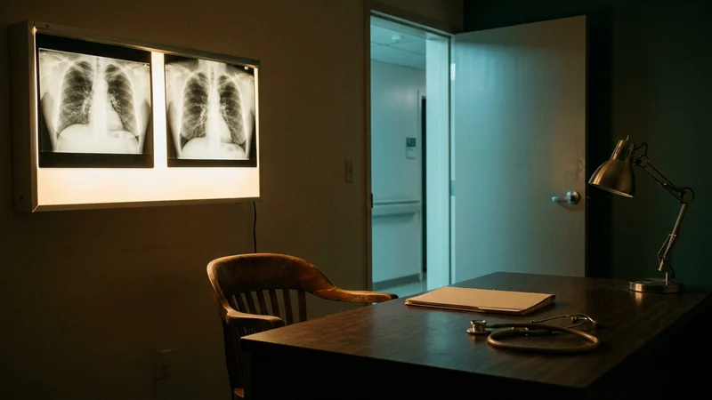
What the Imaging Looks Like
A chest X-ray is the usual first step when someone presents with persistent unilateral chest pain. It is sensitive enough to catch most pleural effusions and many cases of pleural thickening. It is not sensitive for early mesothelioma, because the disease grows as a thin sheet along the pleura rather than as a discrete mass.
CT chest, with contrast, is the diagnostic modality that actually changes the differential. The findings that separate mesothelioma from the other entities on this list are well described:
- Mesothelioma: unilateral pleural effusion, nodular or rind-like pleural thickening (often more than 1 cm), pleural plaques (focal, often calcified), encasement of the lung, and signs of chest-wall invasion in advanced disease.
- Pneumonia: parenchymal consolidation with air bronchograms, with the pleura largely uninvolved.
- Pulmonary embolism: filling defects in the pulmonary arteries on CT pulmonary angiogram, sometimes with wedge-shaped peripheral infarcts.
- Cardiac ischemia: lungs are typically clear on CT; the diagnosis is established with ECG and troponin, not chest imaging.
- Costochondritis: normal CT chest. The diagnosis is clinical, made on examination.
- Rib fracture: focal rib discontinuity on CT, often with a small adjacent pleural effusion (hemothorax) if recent.
Diagnosis of mesothelioma is confirmed by pleural biopsy, not by imaging alone. But the CT findings are usually what move a person from “recurrent unilateral pleural effusion of unknown cause” into a thoracic workup that includes pleural biopsy and immunohistochemistry. People with documented asbestos exposure and pleural thickening on imaging belong in a specialty diagnostic pathway rather than a generic primary-care follow-up.
Why Mesothelioma Pain Becomes Severe in Late Disease
In advanced disease, mesothelioma chest pain often becomes the dominant clinical problem. Bibby and colleagues describe the trajectory: chest pain “tends to worsen as the disease progresses, particularly if invasion of the chest wall occurs,” and “bone pain secondary to rib invasion or neuropathic pain from intercostal nerve involvement may also feature.”
Several mechanisms contribute. The tumor grows along the chest wall and rib cage, producing continuous mechanical pressure on innervated structures. It can erode into ribs, producing focal bone pain. It can encase or compress intercostal nerves, producing neuropathic pain that has a different quality (burning, electric, with allodynia) than the dull ache of the early presentation.
This is why pain control is central to mesothelioma treatment in advanced disease, and why thoracic-oncology programs build pain management into the care plan from the start. A person whose pain shifts from dull and unilateral to constant, severe, and neuropathic has had their disease move into a different phase, and the management has to move with it.
What This Means for Patients and Families
Three practical points fall out of the primary literature:
First, the pattern matters more than the level. A unilateral, dull, persistent ache that does not respond to NSAIDs and is not reproducible on palpation is a different complaint than the central pressure of cardiac angina or the sharp catch of pleurisy. People with documented asbestos exposure should make sure that pattern is described accurately, because that is what places mesothelioma on the differential at all.
Second, the exposure history is the single most useful piece of context. The ATSDR notes that signs of mesothelioma may not appear until 30 to 40 years after asbestos exposure, which is why a clinician seeing a 70-year-old today is most likely seeing the consequences of work that ended in the 1980s or earlier. People should bring up the exposure history explicitly, with details: the industry, the years, the role.
Third, recurrent unilateral pleural effusion is the imaging signal that changes the workup. An effusion that recurs after drainage in someone with chest pain and an asbestos history is an indication for a thoracic evaluation, and when appropriate, a pleural biopsy. A specialty mesothelioma program, such as the Multidisciplinary Mesothelioma Program at the University of Chicago, will set this expectation early, because the time saved by starting the workup at the first recurrence rather than the third is meaningful.
For a fuller picture of how the early symptom pattern tends to evolve, this guide is paired with several others in the same cluster.
References
National Cancer Institute. Mesothelioma Treatment (PDQ)-Patient Version.
https://www.cancer.gov/types/mesothelioma/patient/mesothelioma-treatment-pdq
National Cancer Institute. Mesothelioma Treatment (PDQ)-Health Professional Version.
https://www.cancer.gov/types/mesothelioma/hp/mesothelioma-treatment-pdq
Bibby AC, Tsim S, Kanellakis N, Ball H, Talbot DC, Blyth KG, Maskell NA, Psallidas I. (2016). Malignant pleural mesothelioma: an update on investigation, diagnosis and treatment. Eur Respir Rev. 2016;25(142):472-486..
https://pmc.ncbi.nlm.nih.gov/articles/PMC9487555/
Robinson BW, Lake RA. (2005). Advances in malignant mesothelioma. N Engl J Med. 2005;353(15):1591-1603..
https://www.nejm.org/doi/10.1056/NEJMra050152
Gulati M, Levy PD, Mukherjee D, et al.. (2021). 2021 AHA/ACC/ASE/CHEST/SAEM/SCCT/SCMR Guideline for the Evaluation and Diagnosis of Chest Pain. Circulation. 2021;144(22):e368-e454..
https://www.ahajournals.org/doi/10.1161/CIR.0000000000001029
Proulx AM, Zryd TW. (2009). Costochondritis: diagnosis and treatment. Am Fam Physician. 2009;80(6):617-620..
https://www.aafp.org/pubs/afp/issues/2009/0915/p617.html
Agency for Toxic Substances and Disease Registry. Health Effects of Asbestos Exposure.
https://www.atsdr.cdc.gov/asbestos/health-effects/index.html
Centers for Disease Control and Prevention. Mesothelioma.
https://www.cdc.gov/mesothelioma/about/index.html
Reader Q&A
Frequently Asked Questions
What does mesothelioma chest pain feel like?
Per Bibby and colleagues’ 2016 review in European Respiratory Review, mesothelioma chest pain is usually dull and heavy, sometimes described as a “dragging” sensation. It is most often unilateral, located on the side of the tumor and effusion, and tends to be persistent rather than intermittent. Pleuritic pain (worse with each breath) is less common but can occur when there is parietal pleural irritation.
How is mesothelioma chest pain different from heart attack pain?
The 2021 American Heart Association/American College of Cardiology chest pain guideline describes ischemic cardiac pain as deep, difficult to localize, and usually diffuse, with pressure or tightness in the chest and classic radiation to the arm, jaw, or neck. Mesothelioma pain is unilateral, dull and heavy, and often located along the lower chest wall or back. The AHA guideline notes that pain that is sharp, related to inspiration, or shifts locations, and chest tenderness on palpation, all reduce the likelihood of acute coronary syndrome. ECG and high-sensitivity troponin resolve the cardiac question regardless of the underlying chest-pain pattern.
Is mesothelioma chest pain pleuritic?
Usually not. Bibby et al state that pleuritic pain (sharp pain worse with breathing) is less common in mesothelioma than the dull, heavy, persistent ache. When pleuritic pain does occur in mesothelioma, it is typically due to parietal pleural irritation. The textbook causes of acute pleuritic pain (pneumonia, pulmonary embolism, pneumothorax) usually present more acutely and have distinct imaging signatures.
Can costochondritis be mistaken for mesothelioma?
The two are distinguished by reproducibility on palpation. Per the 2009 American Family Physician review by Proulx and Zryd, costochondritis pain “is reproduced by palpation of the affected cartilage segments and may radiate on the chest wall.” Mesothelioma chest pain comes from the pleura, the effusion, or chest-wall invasion, and it is not reproduced by pressing on the costosternal junctions. An older adult with a known asbestos exposure history whose pain is not reproducible on palpation, and who has a recurrent unilateral pleural effusion, should have mesothelioma on the differential.
Why does mesothelioma chest pain get worse over time?
Bibby and colleagues describe the trajectory: chest pain worsens as the disease progresses, particularly when the tumor invades the chest wall. Late-stage pain often includes a neuropathic component from intercostal nerve involvement and bone pain from rib invasion. Pain control is central to mesothelioma care in advanced disease, and the management has to evolve with the underlying mechanisms.
What imaging is used to evaluate suspected mesothelioma chest pain?
A chest X-ray is the usual first step and can detect most pleural effusions and pleural thickening. CT chest with contrast is the modality that changes the differential, because it shows nodular pleural thickening, pleural plaques, encasement of the lung, and signs of chest-wall invasion. Diagnosis is confirmed by pleural biopsy with immunohistochemistry, not by imaging alone. People with documented asbestos exposure and abnormal pleural findings on CT belong in a thoracic specialty workup rather than a generic primary-care follow-up.
What are the earliest signs of mesothelioma?
Early signs of mesothelioma are often mild and nonspecific, making them easy to overlook. Common early indicators include chest or abdominal pain, persistent cough, shortness of breath, unexplained fatigue, and unintended weight loss. Fluid buildup (pleural effusion in the lungs or ascites in the abdomen) is also frequent in early disease. Because these symptoms resemble common illnesses like pneumonia or irritable bowel syndrome, people with mesothelioma may not receive a diagnosis until the disease has progressed. Early detection is important, as symptoms typically remain mild in stages 1 and 2 but become more severe as cancer advances.
Is mesothelioma terminal?
Mesothelioma is incurable, with no known cure for any stage, and most people with the disease have a median survival of 12-21 months after diagnosis. Stage 4 mesothelioma, the most advanced stage, carries a median survival of about 12 months, though treatments like chemotherapy and immunotherapy can extend life and improve quality of life in some cases. While 12% of people with mesothelioma survive 5 years or more, the disease is generally considered terminal, especially when diagnosed late.